Anthropomorphic 3D-Printed Phantom for Nuclear Imaging Instrumentation

As part of the Freshman Research Initiative in the Advanced Radiological Imaging & Instrumentation Lab, I collaborated on the design of an anthropomorphic, 3D-printed imaging phantom to advance research in nuclear medicine imaging.
Project Overview

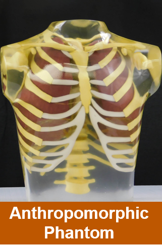
Nuclear medicine techniques such as SPECT and PET require calibration tools called phantoms to ensure imaging accuracy. Commercially available phantoms are often expensive and not adaptable to organ-specific structures. Our project focused on creating a customizable, anatomically realistic, and affordable phantom using digital modeling and 3D printing.
Background

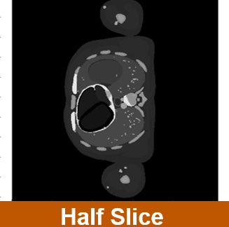

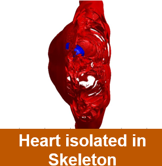

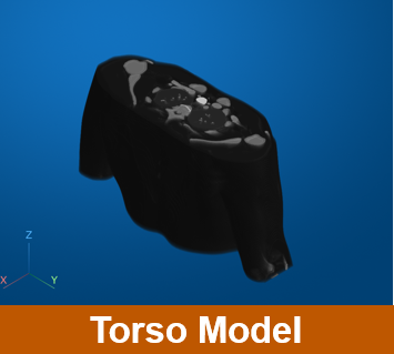
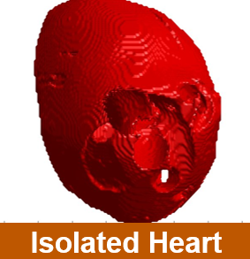


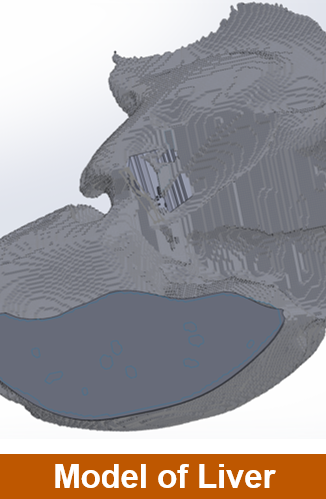
A CT scan of the human torso was imported into MATLAB, containing more than 65 million voxels (3D pixels) across 41 tissue types. From this dataset, I isolated the heart, processed the model to fill voids and smooth surfaces, and prepared it for editing in SolidWorks. The result was a clean digital heart model that could serve as a foundation for future customized phantom development.
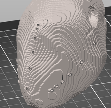
MATLAB Digital Model
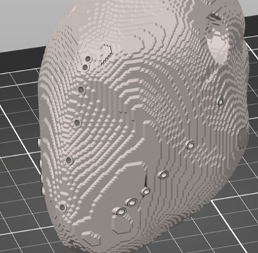
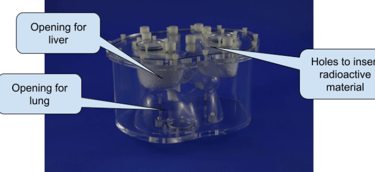
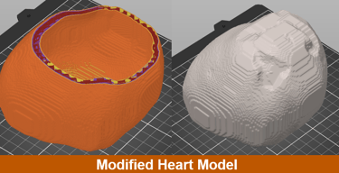
The heart model was exported as an STL file and refined in SolidWorks. I introduced rod channels with diameters of 1.8 mm and 2.8 mm, allowing for the insertion of radioactive sources. These modifications enable the evaluation of spatial resolution and organ-specific disease imaging studies.
CAD Design and Modification
Fabrication
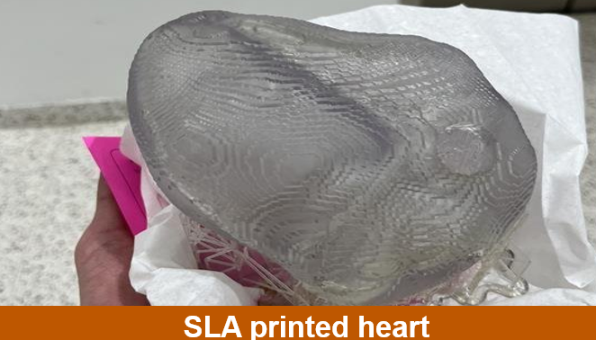
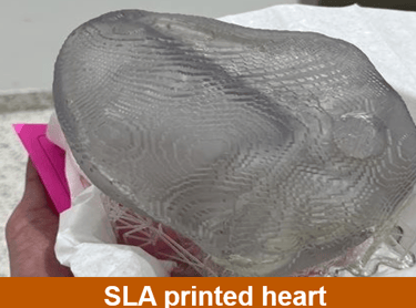
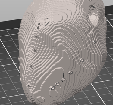
The model was fabricated using SLA resin 3D printing at the Texas Invention Works facility. Printing at a 50-micrometer layer thickness allowed for quality reproduction of fine details. Several test prints were produced, scaled, and adjusted to test accuracy and structural integrity.
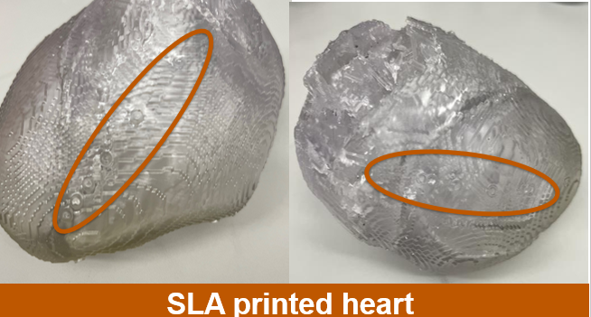
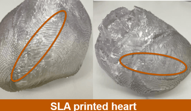
Printed heart phantom with rod holes circled
Future Impact and Work
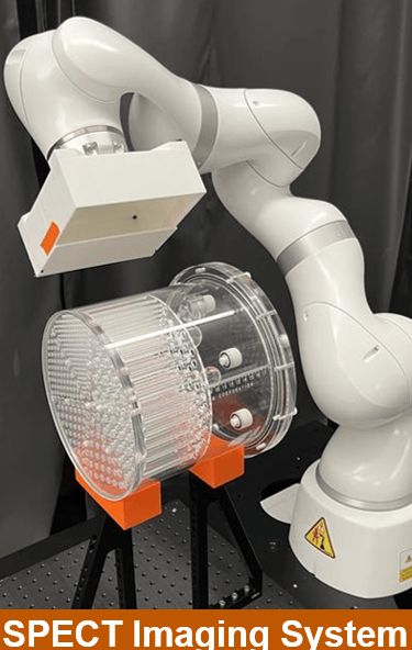
The next stage of this project is to apply the same modeling and printing process to other organs, ultimately assembling a complete torso phantom. Once finalized, the phantom can be tested with SPECT and PET systems to validate its performance in disease-specific imaging tasks. They can then apply this to create patient-specific phantoms through CT scans to give the best imaging testing possible.
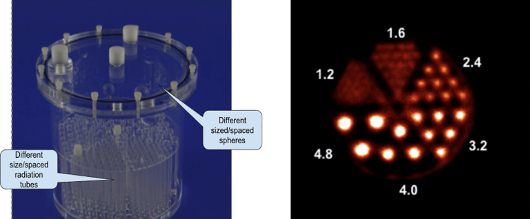
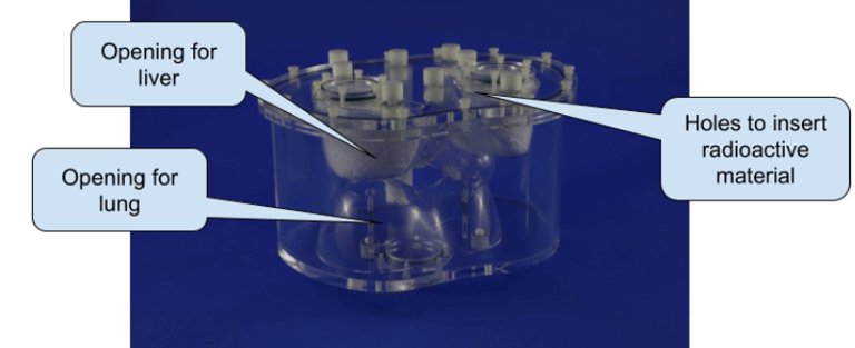
Current Phantoms